Pityriasis rubra pilaris
Downloads
How to Cite
Milano A. 2014. Pityriasis rubra pilaris. Eur. J. Pediat. Dermatol. 24 (1):60 - 61.
pp. 60 - 61
Abstract
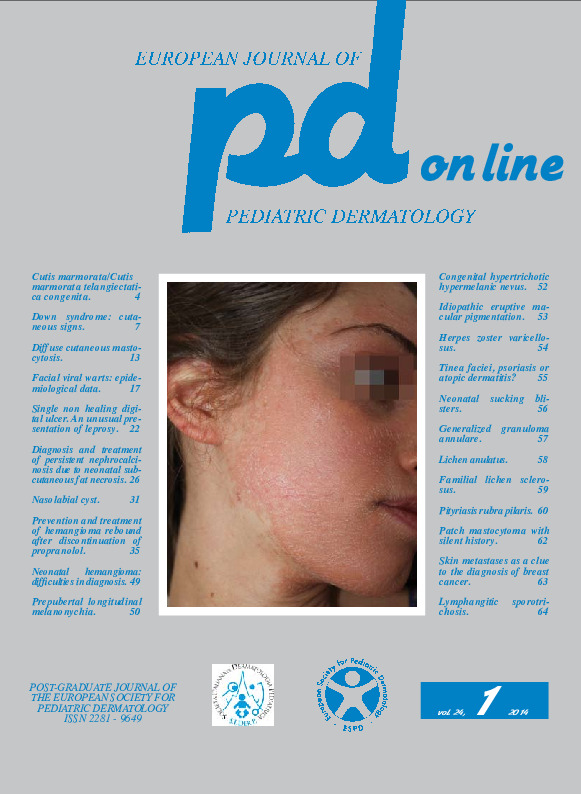
A 17-year-old girl was first observed for the presence of poorly symptomatic, erythematous and scaling lesions that started on the face and then affected the limbs.The mother had anti-thyroid antibodies and she was atopic with allergic rhinitis and sensitized to Dermatophagoides. The physical examination showed confluent erythematous and scaling lesions on the face with clear-cut margins and follicular micropapules at their periphery (Fig. 1); it also showed isolated follicular papules on the limbs (Fig. 5). On the trunk there were modest lesions in correspondence of the hook of the bra (Fig. 4); there was not palmoplantar keratoderma (Fig. 7) and the nails did not show significant alterations (Fig. 8). These clinical features led to the diagnosis of pityriasis rubra pilaris. The lesions responded poorly to the topical therapy, the follicular micropapules of the limbs got confluent to give erythematous and scaling patches with clear-cut borders (Fig. 6) but in the summer regressed completely (Fig. 2).
We reviewed the girl after three years and she told us that the lesions recurred every year during the winter but regressed in the summer. The last winter the recurrence was more severe, affecting always the same locations. The significant involvement of the face (Fig. 3) led us to prescribe acitretin 0.5 mg/kg per day. After 2 months of therapy there was a modest extension of the peripheral lesions and a moderate reduction of the inflammation; recent follicular papules were not visible. From the nosological point of view it is difficult to classify this case, which seems an intermediate form between the type I and II of the adult.
Keywords
Pityriasis rubra pilaris
