Lichen anulatus: usefulness of the dermoscopy
Downloads
How to Cite
Tagliavanti M., Milano A. 2014. Lichen anulatus: usefulness of the dermoscopy. Eur. J. Pediat. Dermatol. 24 (1): 58.
pp. 58
Abstract
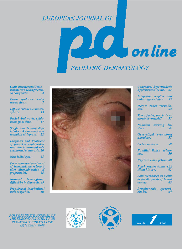
A 16-year-old boy was first observed for the presence of two asymptomatic lesions on the frontal region arisen from 6 months. His family hstory was positive for thyroiditis. The past medical history did not highlight noteworthy diseases. The recent pathological hstory highlighted the appearance of the first lesion in the left frontal region in autumn 2013 and of the second smaller lesion after 2 months.The physical examination (Fig. 1) showed on the frontal region a larger, approximately 12 x 7 mm lesion, well-demarcated, hyperpigmented at the periphery, with irregular lighter areas in the center, a little or nothing infiltrated, and a smaller lesion, which was located in the right frontal region, of about 5 mm in diameter, showing a peripheral, just raised ring. These clinical features were reminiscent of granuloma annulare or lichen anulatus. The dermoscopic examination, showing white amorphous areas with peripheral hypermelanic points led us definitively to the diagnosis of lichen planus anulatus.
Keywords
Lichen anulatus, dermoscopy
