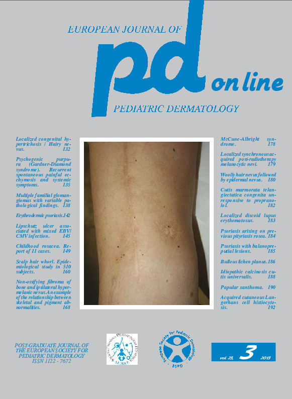
Papular xanthoma in a 6-month-old boy.
Downloads
How to Cite
Bonifazi E. 2013. Papular xanthoma in a 6-month-old boy. Eur. J. Pediat. Dermatol. 23 (3):190-91.
pp. 190-191
Abstract
A 14-month-old child in apparent good health, weighing 11 kg, came to our observation with a history of raised, yellowish, asymptomatic lesions appeared for the first time at the age of 6 months on the back and then gradually increased in number, while the previous lesions resolved spontaneously; the last lesion started at the age of 12 months on the forehead. The physical examination showed on the back a 12 x 7 mm, pink-yellowish nodule (Fig. 1), which became frankly yellow under the pressure of the fingers (Fig. 2). On the back there were also three pink-yellowish papules of about 3 mm in diameter and six pigmentary, sometimes anetodermic residua of variable diameter between 2 and 12 mm. On the right frontal region (Fig. 3) there was a skin-colored papule of 2 mm in diameter. There were no mucosal lesions. The laboratory tests, including cholesterol and triglycerides, were within normal limits. A biopsy was performed with a punch from a papule of the back and the histological examination (Fig. 4, 5) highlighted in the papillary dermis, immediately close to the epidermis, and in the high reticular dermis a monomorphic population of large cells with abundant foamy cytoplasm and central nucleus with evident nucleolus. There were a few histiocytes without Touton cells and inflammatory infiltrate. The diagnosis of papular xanthoma was done and its favorable prognosis with tendency to spontaneous resolution was confirmed to the family.Keywords
Papular xanthoma
