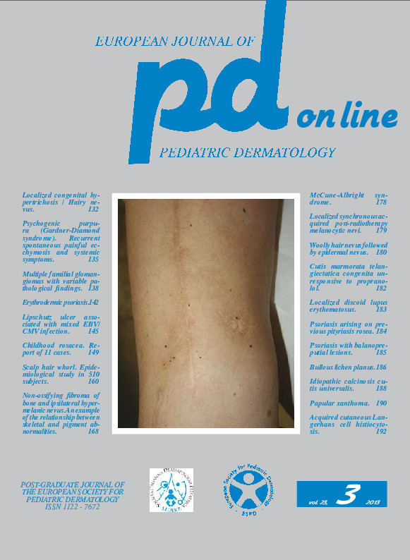
Idiopathic calcinosis cutis universalis.
Downloads
How to Cite
Garofalo L., Milano A. 2013. Idiopathic calcinosis cutis universalis. Eur. J. Pediat. Dermatol. 23 (3):188-89.
pp. 188-189
Abstract
The patient was a first-born, born after 35 weeks of gestation by cesarean section due to maternal heart disease, with a weight of 1,840 g and a length of 44 cm at birth. From the first months of life his parents noticed nodules and plaques of hard consistency especially in the trunk; there was no family history of similar lesions. There were no symptoms referable to a connective tissue. At the age of 3 years he received a diagnosis of autism. The dermatological examination at one year of age showed nodules and plaques of hard consistency, varying in diameter from a few mm to 3 cm in the submental, left subcostal and left inframammary region, above the right iliac crest, in the low back, on the extensor surfaces of both forearms, right buttock and right popliteal fossa. The larger lesion was in the left subcostal region: it was a plate of 3 cm in diameter, a couple of millimeters thick, which could be stapled with the hand (Fig. 1, 2). The laboratory tests showed microcytosis, serum calcium 5.3 mEq/L (n.v. 4.5-5.5), phosphoremia 6.06 mg/dl (n.v. 4-6, 5), parathyroid hormone 64.05 pg/ml (n.v. 7-83 ), urinary phosphorus 0.12 g/24h (n.v. 0.5-0.8), tubular reabsorption of phosphate 85% (n.v. 80-90), U- deoxypyridinoline 23.5 nM/mM (n.v. 1.5-7), osteocalcin 26 ng/ml (n.v. 5-18), somatomedin 47.6 ng/ml (n.v. 50-426). There were no autoantibodies and CPK and transaminase levels were within normal levels. Molecular analysis of the GNAS gene showed no abnormalities. The histological examination of a skin lesion showed calcium deposits in the dermis and subcutaneous fat tissue (Fig. 3, 4). These laboratory findings led to the diagnosis of idiopathic calcinosis cutis universalis. The minor esthetic relevance of the lesions and the absence of subjective symptoms led us to a wait and see policy. Over the next six years of follow- up there were no significant changes compared to the first observation, the left subcostal plaque remained unchanged (Fig. 6), modest scars were noticed on the back (Fig. 5 ) as residua of ulceration with leaking of chalky white material from the lesions.Keywords
Idiopathic calcinosis cutis

