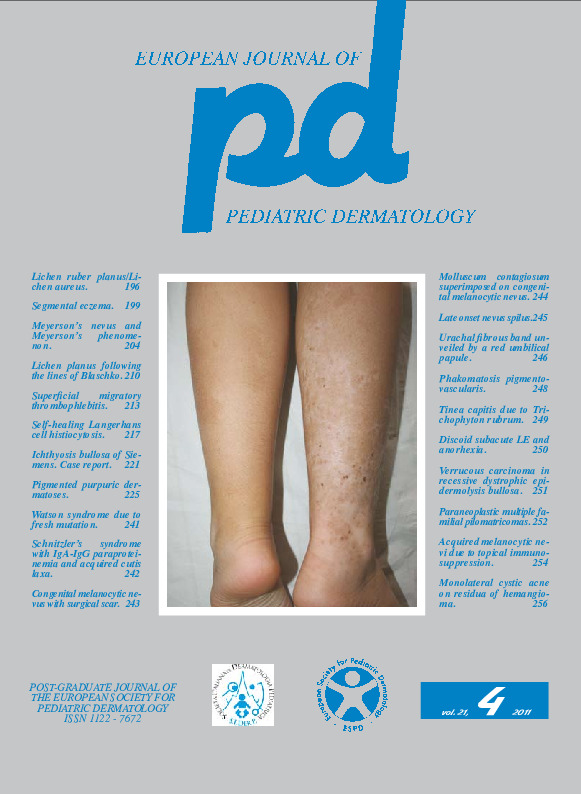
Phakomatosis pigmento-vascularis.
Downloads
How to Cite
Garofalo L. 2011. Phakomatosis pigmento-vascularis. Eur. J. Pediat. Dermatol. 21 (4): 248.
pp. 248
Abstract
Case report. A 9-year-old, red-haired girl presented hyperpigmentaion throughout the skin surface since birth. Physical examination revealed lesions of two types: on the abdomen bilaterally (Fig. 1) and lower limbs, predominantly on the left, there were asymptomatic, uniform in color brownish spots, with clear-cut borders and reticular dermoscopic pattern (Fig 1). There were also 5-8cm in diameter, telangiectasic patches in the left retroauricular (Fig. 3) and gluteal region; moreover, there were 0.5-1 cm in diameter, isolated telangiectasias on the trunk and limbs (Fig. 2 circled). Telangiectasias disappeared under finger pressure and were well visible dermoscopically (inset Fig. 3). Histology of the retroauricular region showed dilated capillaries in the superficial dermis (Fig. 4), while the brown spots showed increased melanin in the epidermis. Phakomatosis pigmentovascularis was the final diagnosis.Keywords
Phacomatosis pigmentovascularis

