Acquired self-healing cutaneous Langerhans cell histiocytosis.
Downloads
How to Cite
Mazzotta F., Bonifazi E. 2010. Acquired self-healing cutaneous Langerhans cell histiocytosis. Eur. J. Pediat. Dermatol. 20 (3):214-15.
pp. 214-215
Abstract
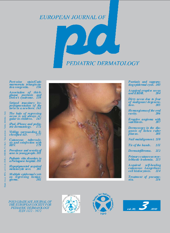
Case report. A little girl followed in our clinic from the age of 3 months for a deep hemangioma of the right scapular region at a control visit performed at 1 year presented papules of the diaper region (Fig. 4), on the skin of the hemangioma (Fig. 1 and inset) and shortly after on the scalp (Fig. 2), in the axillary region and scattered on the anterior chest (Fig. 3 and inset), and finally in the retroauricolar region (Fig. 6). We were dealing with 1-2 mm in size, scaling, non-bleeding, isolated, monomorphic papules; in the right scapular region below the papules could be seen the bluish discoloration of hemangioma (Fig. 1), which appeared at palpation like a mass of 3 cm (inset of Fig. 1). The histological examination of a papule showed an infiltrate composed of large vesicular histiocytic cells, with kidney-shaped nucleus and large acidophil cytoplasm. On immunohistochemistry these cells were S-100, CD1a, CD68 positive leading to the final diagnosis of Langerhans cell histiocytosis. The patient was sent to the oncohaematologists. X-ray of the skeleton did not highlight osteolytic images, routine tests were within normal limits, abdominal ultrasound did not show alterations of the hypochondriac organs and a bone marrow biopsy showed normal cellularity. The final diagnosis was isolated cutaneous Langerhans cells histiocytosis and the child entered a protocol of clinical and laboratory monitoring which provided a monthly skin and oncohematology examination, with blood counts and abdominal ultrasound. For 8 months the child continued to present subintrant eruptions of asymptomatic micro-papules in the same locations (Fig. 5, 6). The individual lesions regressed within a few weeks. Changes in clinical or laboratory findings tending to show an involvement of other organs were never noticed. After 9 months no new lesions appeared and at the age of 22 months there were only discolored residua (Fig. 7, 8). During the subsequent 6 months the little girl, checked every two months now, no longer presented skin lesions, leading to the final diagnosis of self-healing cutaneous Langerhans cell histiocytosis.Keywords
Langerhans cell histiocytosis
