Pigmented epithelioid melanocytoma: a pediatric case.
Downloads
DOI:
https://doi.org/10.26326/2281-9649.35.1.2725How to Cite
El Bouhmadi A., El Fatoiki F., Diouri M., Hali F., Chiheb S. 2025. Pigmented epithelioid melanocytoma: a pediatric case. Eur. J. Pediat. Dermatol. 35 (1):27-9. 10.26326/2281-9649.35.1.2725.
pp. 27-9
Abstract
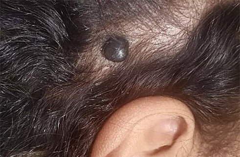
Pigmented epithelioid melanocytoma (PEM) is a rare, low-grade melanocytic tumor that primarily affects children and young adults. This report details a case of an 8-year-old girl presenting with a pigmented scalp lesion. Clinical examination revealed a black nodular tumor, confirmed through dermoscopy and histopathology to be PEM, characterized by large, pigmented melanocytes with epithelioid or spindle-shaped morphology. Immunohistochemistry showed positive staining for Melan-A and HMB-45. The lesion was surgically excised with no lymph node involvement or metastasis on follow-up. Despite potential regional lymph node metastasis, PEM typically has a benign clinical course, and complete surgical excision remains the preferred treatment.
