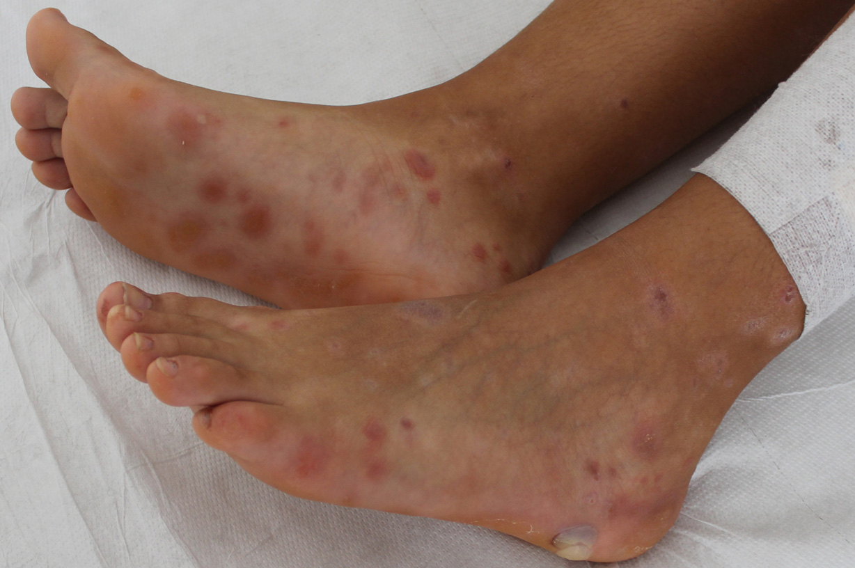
Non-bullous recurrence of bullous lichen planus after 8 years of well-being.

Downloads
DOI:
https://doi.org/10.26326/2281-9649.32.1.2323How to Cite
Abstract
Lichen planus (LP) is rare in children and bullous lichen planus (BLP) is even more exceptional (1, 2, 4, 5). BLP, as the current case demonstrates, is not a disease in its own right, but is only a clinical variant of LP characterized by the presence of blisters that appear exclusively on pre-existing papular or erythematous-purple lesions of lichen planus. It is likely that the blisters are favored in their onset both by the vacuolar degenerative phenomena of the basal layer of the epidermis, and by the massive banded lymphocytic infiltrate in the superficial dermis.
Little is known about the causes of bullous lichen planus. There are some familial cases in the literature (3), which would be transmitted in an autosomal dominant way with variable penetrance; compared to unfamilial cases, they are characterized by a more extensive involvement of the skin and an earlier period of onset. Among the exogenous causes of LP vaccination, in particular against hepatitis B, is reported in 18 cases in the literature (6) and surprisingly in this report as many as 5 out of 18 cases are of bullous lichen planus, while in a large series of 87 cases of childhood lichen planus only one case of bullous lichen planus is reported (2).
The presence of blisters requires differential diagnosis from other autoimmune bullous skin disorders, especially bullous pemphigoid, bullous lupus erythematosus, and lichen planus pemphigoides. Dermatological examination is fundamental and is based on the identification of classic LP lesions, especially the flat surface papules and pigmentary outcomes, which allow to exclude bullous pemphigoid and lupus erythematosus. Differential diagnosis from lichen planus pemphigoides, in which bullous lesions and lichen papules coexist, is more difficult, but blisters also form on healthy skin not affected by lichen. Immunofluorescence facilitates the differential diagnosis from lichen planus pemphigoides, as well as from bullous pemphigoid and lupus erythematosus.
