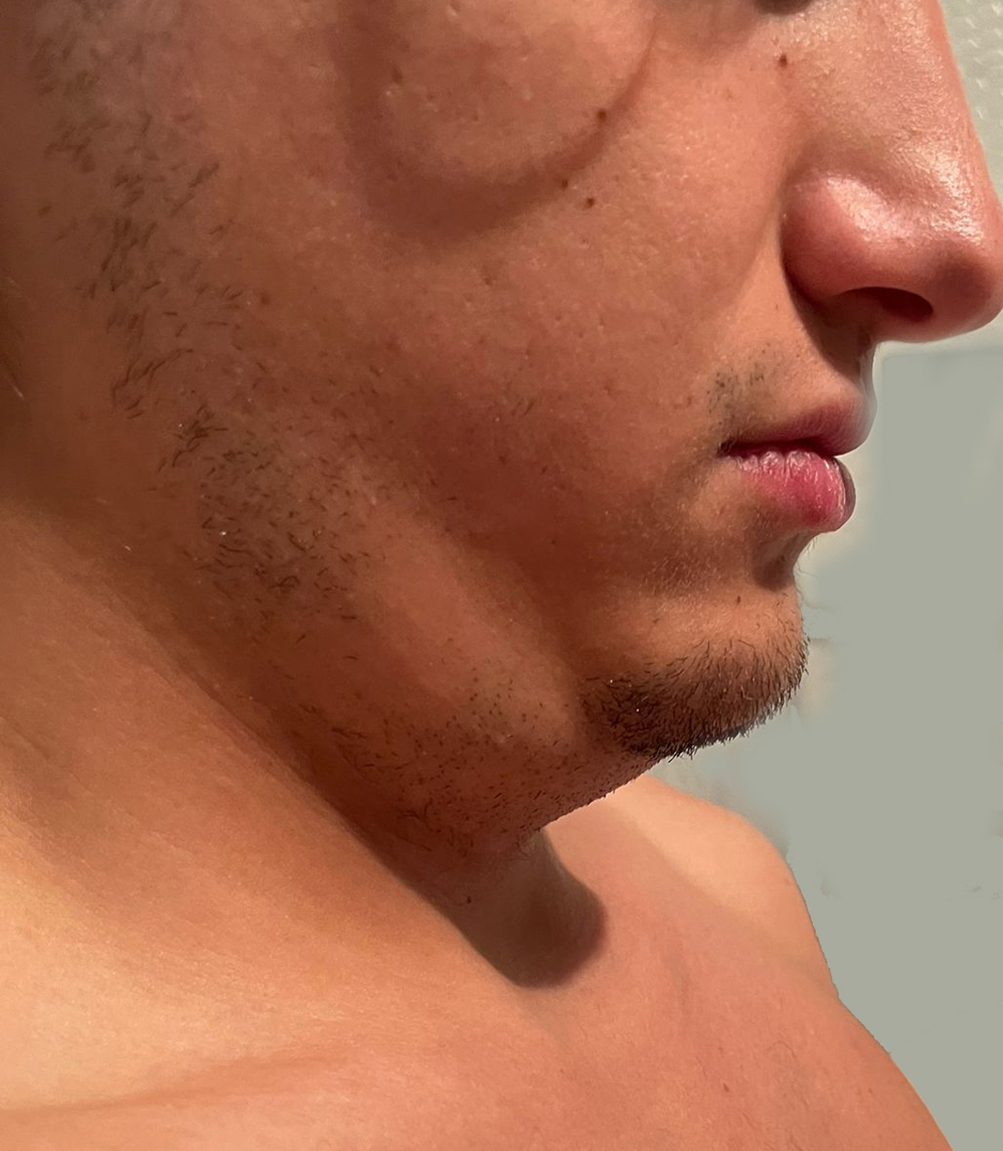
Diving ranula.

Downloads
DOI:
https://doi.org/10.26326/2281-9649.31.4.2296How to Cite
Abstract
The diving ranula is a collection of extravasated saliva that originates from a salivary gland, usually sublingual, more rarely submandibular or ectopic; often a small defect of the mylohyoid muscle favors the extravasation of fluid into the submandibular space; the cavity which contains the liquid has no epithelial lining and is made up of fibrous connective tissue (4), so it is a pseudocyst. Its annual incidence is 2.4 per 100,000 subjects, it slightly prevails in males and is usually diagnosed in the third decade of life (1).
The pathogenesis is unknown. However, trauma, inflammatory phenomena, occlusion of the glandular excretory ducts have been hypothesized; genetic malformative factors, for example a hiatus of the mylohyoid muscle are also possible, as seems to confirm an increased incidence in some ethnic groups (1).
The differential diagnosis of diving ranula is difficult because it must be done with all neck swellings, usually benign in young people (2, 6), but especially with macrocystic lymphangioma and branchial cyst, which have a liquid content, are compressible and present volumetric fluctuations like diving ranula. Lymphangioma is the most frequent of these three disorders. It generally begins in the first years of life, is located in the posterior triangle of the neck behind the sternocleidomastoid muscle and typically undergoes intralesional hemorrhages that make it become much larger and can transpire on the surface with the characteristics color changes of bruising. Branchial cysts are found on the anterior border of the sternocleidomastoid muscle and frequently undergo infection. In case of doubt, the aspiration of the liquid highlights a high content of amylase in diving ranula (3).
The natural history of diving ranula is not known but even in the event of a progressive increase due to its liquid content and the anatomical characteristics of the neck, compression phenomena of the vessels, airways or pharynx can hardly occur. Its spontaneous regression or following modest inflammatory reactions is also possible (8).
