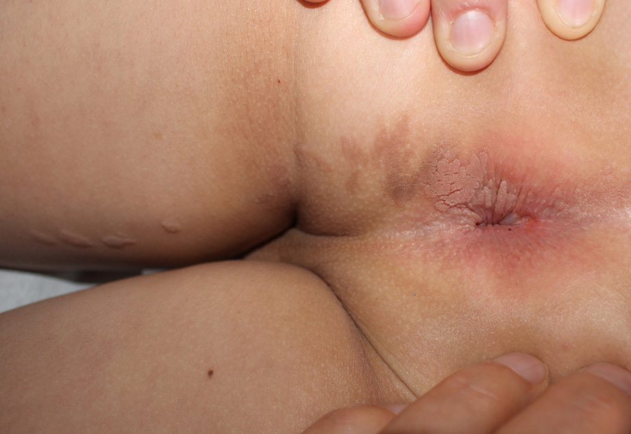
Verrucous epidermal nevus: diagnostic difficulties.

Downloads
DOI:
https://doi.org/10.26326/2281-9649.31.4.2287How to Cite
Abstract
Along Blachko’s lines are distributed diseases with Mendelian inheritance such as incontinentia pigmenti, polygenic hereditary diseases such as psoriasis or atopic dermatitis (3), nevi such as verrucous epidermal nevus (VEN) and its inflammatory variant (1), and acquired diseases such as lichen striatus (LS). All these diseases share a condition of mosaicism, that is, they presuppose the existence of a gene mutation, often post-zygotic, that gives rise to a clone of cells different from all the others. The type of mutated gene is responsible for the different morphology of these diseases, but the distribution on the skin of the mutated clone is always the same; therefore, it does not depend on the gene, but on the time in which the mutation occurred and on the stereotyped movements of the migrations that skin cells undergo during fetal life. If the nevus is very extensive and therefore secondary to an early mutation, other organs may be involved such as the nervous system, the eye, the skeletal system, as happening in epidermal nevus syndrome (4) or even some gonadal cells with the risk that the mutation is transmitted to the zygote of the offspring thus giving rise to a generalized inherited disease (5).
VEN is usually present at birth; when it is localized in hidden locations or when it initially manifests with just hyperpigmented lesions it may become evident later. However, it is exceptional that it manifests itself at 8 years as in the present case. Its earliness and its morphology are able in most cases to easily differentiate it from an acquired inflammatory condition such as LS.
LS is a self-healing inflammatory skin disease, usually asymptomatic, which affects the first years of age, usually between 5 and 15 years, and is distributed linearly along Blaschko’s lines, suggesting that on the line in which it occurs there is a clone of mutated cells as a result of a post-zygotic mutation: this mutation would give the cells involved a particular susceptibility towards an exogenous agent that, upon its arrival, would cause the transient inflammatory reaction (2). There are various hypotheses on the pathogenetic mechanisms that lead to the inflammatory reaction and its spontaneous regression.
The present case has posed problems of differential diagnosis between VEN and LS. The histological findings were not able to eliminate the clinical doubt, indeed they added another possible differential diagnosis, i.e. inflammatory linear verrucous epidermal nevus. The latter diagnosis was discarded due to the lack of itching and obvious clinical signs of inflammation. Only prolonged clinical observation over time resolved the diagnostic doubt thanks to the persistence of the lesions and the greater evidence of papillomatous and warty structures.
