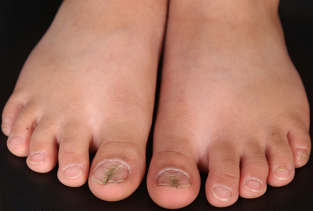
Bilateral median canaliform dystrophy of great toenails in a child.
Downloads
DOI:
https://doi.org/10.26326/2281-9649.31.4.2286How to Cite
Abstract
Median canaliform dystrophy is a rare form of nail abnormality, first described by Heller in 1928 (5). It usually involves one or both thumbnails as a midline longitudinal split with multiple transverse lines radiating from the center, resembling a fir tree (5).
Toenails or other fingernails are rarely affected (4, 5). There are several reports of median canaliform nail dystrophy occurring after isotretinoin treatment of acne vulgaris (1, 3) and one case report after cryotherapy of periungual warts (2). Although minor trauma to the nail matrix has been hypothesized as an etiology, most cases are idiopathic, and patients could not identify any preceding trauma (2). Familial cases have been reported, but the role of genetic factors in these cases is not well established (5).
Median canaliform nail dystrophy should be differentiated from habit tic deformity, in which the patient admits manipulation of the periungual area when questioned appropriately (5). Our patient and his parents denied any habitual picking or trauma to toenails. The involvement of both great toenails and the onset in early infancy made habit tic deformity less probable.
Median canaliform nail dystrophy occasionally resolves spontaneously within several months to years (3, 5). The patient should avoid any minor trauma to the nail plate and the periungual area. It is recommended to keep nails short and prevent any nail plate catching by covering it with either tape or a nail wrap (5). Topical corticosteroids or tacrolimus 0.1% under occlusion are other treatment options, but their effectiveness must be elucidated (1).
