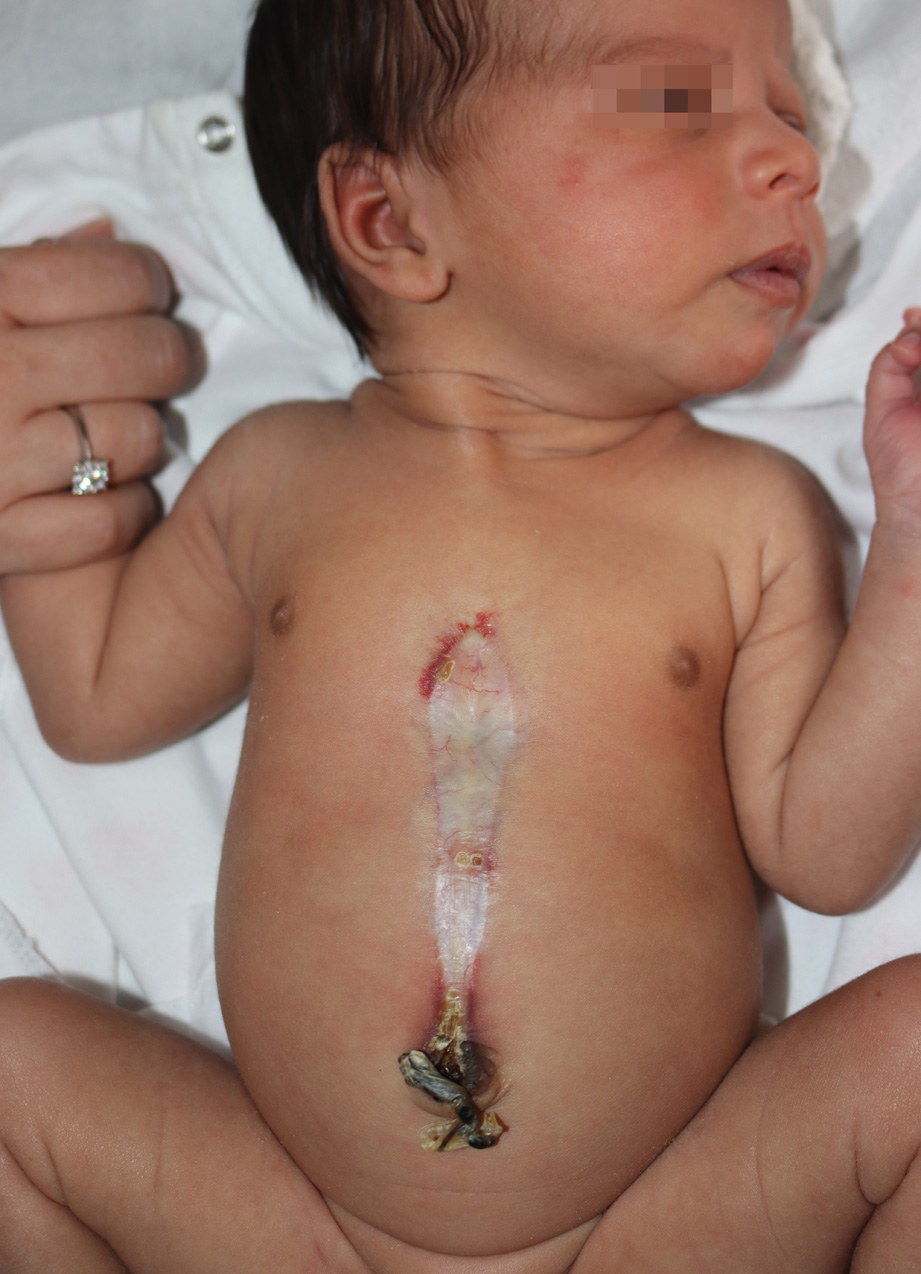
Supraumbilical midline raphe and infantile hemangioma.
Downloads
DOI:
https://doi.org/10.26326/2281-9649.31.3.2268How to Cite
Abstract
Midline skin malformations are very common in the newborn; in most cases, the latter are isolated skin lesions of little clinical importance; on the other hand, in some cases they are a clue to the diagnosis of possible malformations of the underlying tissues with severe prognosis.
An example of this second occurrence is the supraumbilical raphe. Closure on the midline occurs around the eighth week of gestation when the mesoderm merges on the midline. When this mesodermal fusion does not occur, the overlying ectodermal epithelium undergoes necrosis. The supraumbilical raphe is believed to result from this lack of midline ecto-mesodermal interaction, followed by scar repair (6).
The umbilical raphe can be an isolated skin defect or associated with sternal cleft, Cantrell pentalogy with cardiac malformations up to ectopia cordis, vascular dysplastic syndrome and PHACES syndrome (4). The supraumbilical raphe can be associated with hemangiomas in the absence of other malformations (2). However, other malformations are more frequently present, as in PHACES syndrome. In the latter case, the umbilical raphe can represent the first and only sign at birth; hemangioma can in fact manifest itself after days or weeks, while posterior fossa malformations and encephalic and cervical vascular dysplasias can be only unveiled with imaging techniques; if ignored, vascular dysplasias can be unveiled even after decades by stroke episodes (1).
These cases reopen the discussion of the relationship between malformations and hemangiomas. Although usually independent, it is possible that vascular malformations may be responsible for hemangioma: in these cases the hemangioma could be the expression of an exaggerated angiogenic response to the condition of hypoxia caused by vascular dysplasia (5). However, it is also possible that the same pathogenetic factor, maybe of maternal origin, acting at different times, could be responsible for both early malformations, which occur during the first three months of gestation, and hemangiomas, which occur in the perinatal period. There are data from the literature (3) that really show the presence in the same family of limb defects secondary to vascular malformations in some relatives and hemangiomas in others.
The actual case series confirms that midline supraumbilical raphe and hemangioma may be present in the same individual as in the first case. However, it also emphasizes that the two disorders can occur separately in two twins.
