Follicular mucinosis in an 11-year-old boy.
Downloads
DOI:
https://doi.org/10.26326/2281-9649.31.1.2217How to Cite
Abstract
Discussion. Follicular mucinosis (FM) is a histological term which means accumulation of mucin, i.e. mucopolysaccharides or glucosaminoglycans, in the outer sheath of the hair follicle and in the sebaceous gland; this histological picture can be found, in addition to idiopathic FM, in numerous benign conditions, such as lupus erythematosus (8), eosinophilic folliculitis (4), epidermal nevi (7), leprosy (3), and malignant disorders as follicular lymphomatoid papulosis (6) and especially folliculotropic mycosis fungoides (FMYF). The equivalent term of alopecia mucinosa, which introduces a clinical sign – alopecia – seems more appropriate to define the disease. However, alopecia is not always evident, especially when is not affected an area of skin with terminal hair.
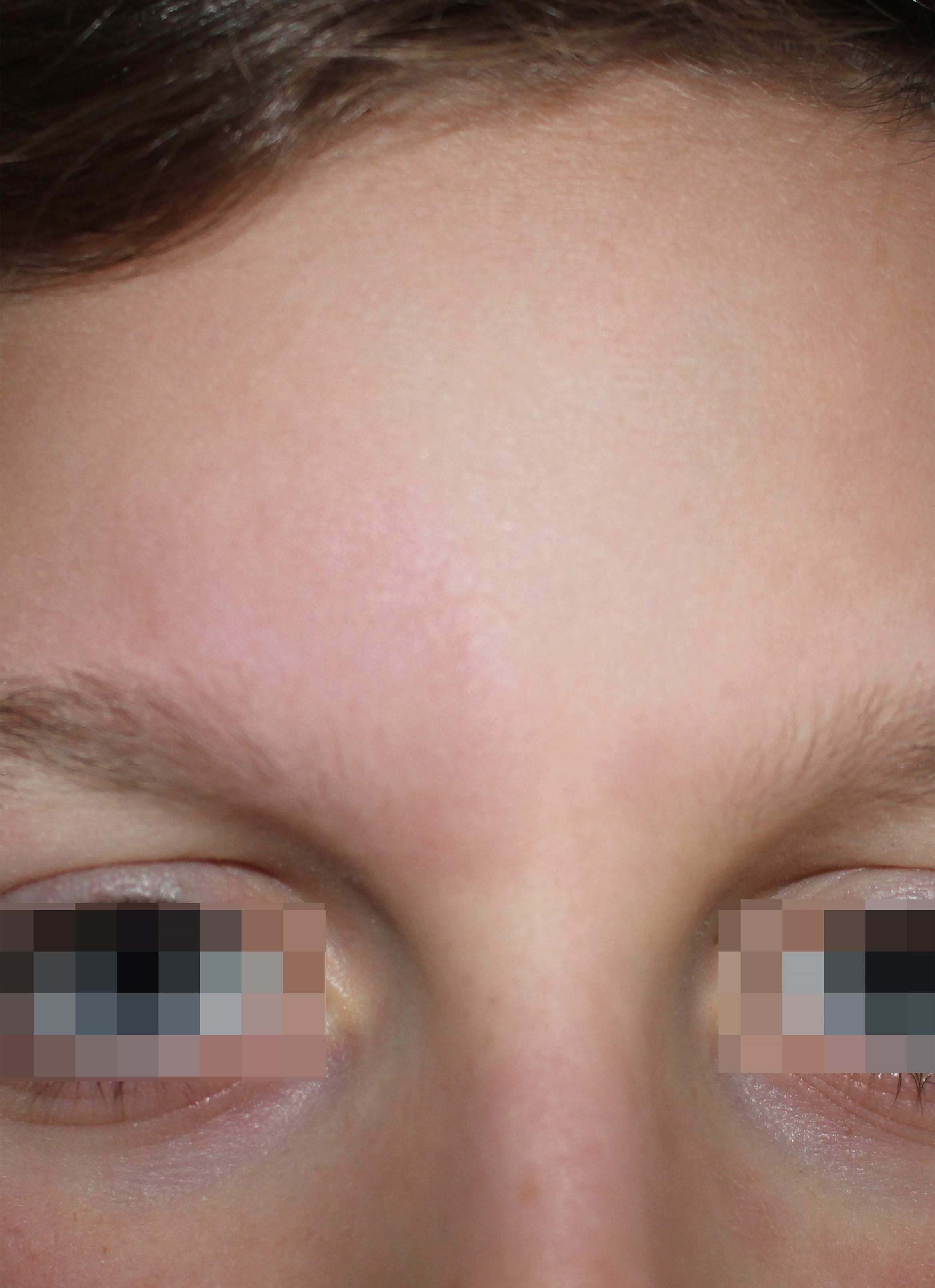
Follicular mucinosis presents clinically with clustered skin-colored papules or erythematous, sometimes scaling plaques, in which follicular reliefs may be evident. Based on the clinical course, we distinguish a form with one or a few lesions that resolve spontaneously within a few months, a form with more numerous elements, of more variable morphology that recur for many years and a form associated with lymphoma, generally with mycosis fungoides.
The most important clinical problem is the distinction between idiopathic FM and FM associated with lymphoma, especially mycosis fungoides (1, 8). FMYF is the most frequent lymphoma associated with FM. However, the frequency of the association varies according to the different Authors between 9.4 and 64% of all cases of FM (1). Although FMYF can occur several years after the diagnosis of FM, in most cases FM and FMYF occur simultaneously (1). With regard to the association MF-FMYF children and more generally young patients and female subjects are less likely to have an associated FMYF (5). Most Authors agree that the onset of FM in the head with one or a few lesions has a benign clinical course (2). Monoclonality and TCR gene rearrangement, although more frequent in FMYF-associated forms of FM, do not represent a sure sign of malignancy (1).
The present case was presented for its rarity; the presence of a single head lesion in a young boy and the polyclonal nature of the infiltrate speak in favor of a benign course, not excluding the need for careful clinical monitoring.
