Atypical vascular proliferation in chronic radiodermatitis secondary to radiotherapy for infantile hemangioma.
Downloads
DOI:
https://doi.org/10.26326/2281-9649.31.1.2213How to Cite
Abstract
Radiotherapy is still used today in the treatment of infantile hemangioma (IH) in the case of particular localizations such as the pituitary fossa, when other treatments are not possible (1). On the other hand, it is not actually indicated in cutaneous IH, even if it was still used until the ’60s.
Among its side effects there is chronic radiodermatitis (6), which today is observed mainly in women undergoing radiotherapy for breast cancer.
The possibility of developing radiodermatitis depends on various factors, including the proximity to the skin of the radiotherapy target, the radiation energy, the overall dose of radiation and its treatment scheme, and the size of the skin surface exposed to radiation.
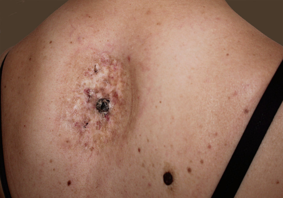
Chronic radiodermatitis is histologically characterized by fibrosis of the dermis with disappearance of the hair follicles and glands, and rarefaction of the vessels; some residual vessels can be very dilated. The epidermis can be acanthotic or atrophic and characterized by numerous dyskeratotic cells.
However, a more fearful side effect of radiotherapy for IH is the late onset, even after 6 decades (8) of tumors (2, 3, 7), including angiosarcoma (5) and melanoma (4).
Besides malignant tumors there are also benign proliferations. The atypical vascular proliferations on radiodermatitis are benign tumors consisting of branched and dilated vessels or irregular vascular lacunae delimited by endothelium; degeneration into angiosarcoma has been described in 3-6% of cases (8). The considerable delay between irradiation and the appearance of these proliferations seems to be linked to the low doses of X-rays used in the treatment of hemangioma (8).
The current case was presented due to the rarity of atypical vascular proliferations and to remind us that neoformations on radiodermatitis are not always malignant.
