Angioma as a clue to the diagnosis of xeroderma pigmentosum.
Downloads
DOI:
https://doi.org/10.26326/2281-9649.30.3.2142How to Cite
Abstract
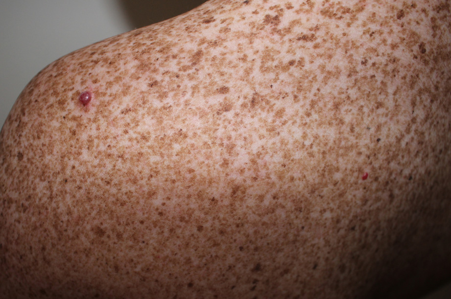
Normally, the diagnosis of xeroderma pigmentosum (XP) becomes clear due to the early onset of non-melanocytic skin tumors (NMT) and melanoma (M). Due to the well-known unable excision-repair of nucleo-tides damaged by ultraviolet rays, subjects with xeroderma pigmentosum have a 10,000 times higher risk of NMT; the risk of M is lower but still 2000 times greater than normal subjects. This fact makes us reflect on the different role of RUVs in the genesis of NMT and M. Even more it makes us reflect the fact that while in the normal subject the average age of onset of M is earlier than that of NMT, in the subject affected by XP the age of onset of melanoma is later than that of NMT (2). In the current case the diagnosis was made thanks to the onset of two angiomas at 10 years of age. We have not found in the literature cases of xeroderma pigmentosum (XP) that have come to observation and have been diagnosed as such due to the presence of angiomas; there are descriptions of angiomas (3), epithelioid angioma (5), cavernous hemangioma (4), angiokeratoma (1) but these angiomas occurred in subjects who had already received a diagnosis of XP. In the present case, the initial diagnostic indication of extreme sensitivity to RUV despite the patient’s dark phototype was not understood and surprisingly the early and profuse appearance of freckles in photo-exposed sites passed over in silence, because it was given the same meaning as the sunburns of the father and his rutile relatives.
