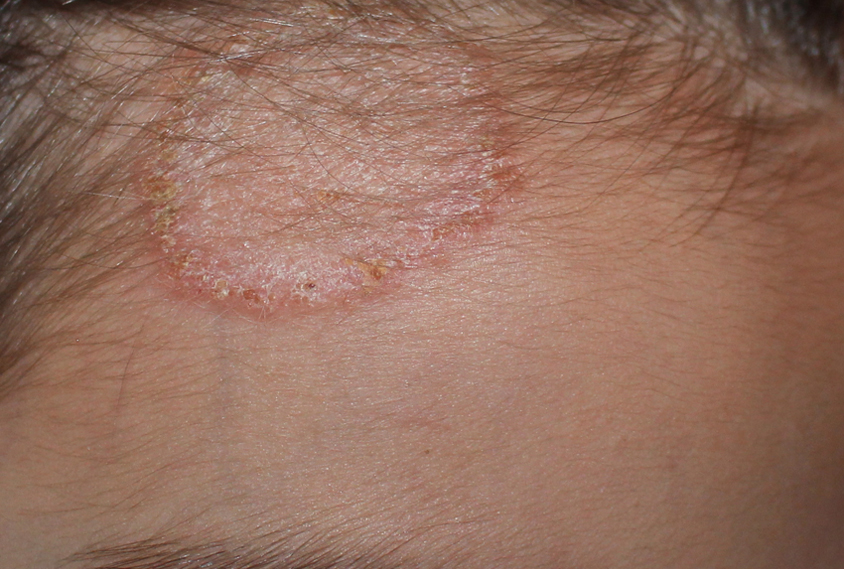
Frontal tinea corporis followed by tinea capitis.
Downloads
DOI:
https://doi.org/10.26326/2281-9649.30.1.2083How to Cite
Abstract
The current case is interesting due to some therapeutic and pathogenetic implications. At the time of the first examination, only tinea corporis of the frontal region was present and hair involvement was not clinically evident. Nonetheless, it was decided to start griseofulvin orally because of the proximity of the mycotic infection to the hair-line (1). The presence of truncated and parasitized hairs after 2 weeks despite oral antimycotic therapy can be easily explained if the pathogenetic mechanisms of tinea capitis - well highlighted by Kligman’s experimental studies - are taken into account (2). The fungi coming from the outside penetrate deeply into the hair follicle up to the level of the bulb and the hair infection begins 1 mm above the bulb; subsequently the infection progresses distally at a rate of about 0.3 mm per day reaching the follicular ostium after about 2 weeks and then causing the hair to break.
