Focal congenital lipoatrophy: minimal growth hemangioma or malformation?
Downloads
DOI:
https://doi.org/10.26326/2281-9649.29.4.2050How to Cite
Abstract
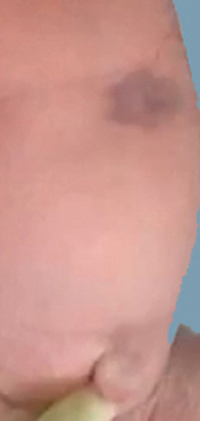
A focal congenital lipoatrophy has been described concomitantly with vascular lesions, both in minimal growth reticular hemangioma (1) and in vascular malformations (7). In the first report (1) lipoatrophy was associated with unequivocal signs of hemangioma such as reticulated telangiectases, ischemic halo and above all hemangiomatous proliferative papules. In the second report (7) lipoatrophy was not associated with telangiectases and proliferating papules; the histology spoke of a capillary malformation confirmed by the negativity of GLUT-1 in endothelial cells.
Lipoatrophy could be a trait d’union between hemangioma and vascular malformations; lipoatrophy and hemangioma could both be related to hypoxia which inhibits adipocyte differentiation and stimulates angiogenesis (1); hypoxia could sometimes be the consequence of a vessel malformation.
The following observations support the hypothesis that hypoxia can sometimes be secondary to a vessel malformation: in case 1 of the report of Bessis et Al. (1) focal lipoatrophy was present in a minimal growth hemangioma of the thigh that underwent an unusual ulceration both for the site, outside the diaper, and above all for the time of onset, at the seventh month of life. In 2008 Mazzotta et Al. (3) described 3 cases of minimal growth, segmental hemangiomas of the lower limbs that underwent ulceration at the age of 7-8 month and in one of these cases documented a malformation of the iliac artery as responsible for ulceration. Other Authors stressed the relationship between minimal growth hemangiomas, ulcerations and arterial malformations, such as ectasia of the common iliac and hypoplasia of the femoral artery (4). The relationship between hypoxia due to vascular malformation and hemangioma has been repeatedly reported in the literature, especially but not only in the PHACE, PELVIS, SACRAL syndromes; you should also consider the hemangiomas associated with Poland syndrome (6), with the absence of the carotid artery (5), and with vascular anomalies responsible for acral limb defects (2). These reports confirmed the possibility that a vessel malformation may be responsible for hypoxia: hypoxia in turn could determine both hemangioma by stimulating reactive angiogenesis and sometimes lipoatrophy by inhibiting lipogenesis.
