Pseudobullous aplasia cutis with dyspraxia.
Downloads
DOI:
https://doi.org/10.26326/2281-9649.28.3.1886How to Cite
Abstract
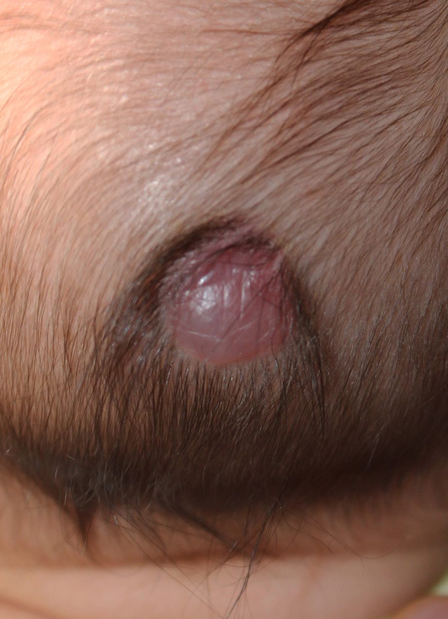
Congenital aplasia cutis is due to a defect in closing the skin. Typically localized to the scalp, it occurs at birth as a very extensive, deeply ulcerated lesion with possible involvement of the cranial plate and the meninges or more frequently as a single or multiple lesion, roundish, of 1-3 cm in diameter, with definite borders and without hair. The small lesion can be eroded, ulcerated or already scarred; the scar, which is already present at birth or subsequent, can be atrophic, normotrophic or hypertrophic; the lesion of small dimensions can have an atrophic, bright, membranous-like surface (membranous aplasia cutis); it can be surrounded by a thicker, more pigmented hair ring and can resemble a blister.
However, it is not actually a blister because it does not erode and does not form a crust (1, 2): the pseudo-bullous lesion is whitish, greyish or bluish, its surface may be taut or creasy; at palpation it is characteristically soft-elastic, sometimes painful; finally it can be surrounded by a thicker and darker hair (5).
The clinical features of pseudo-bullous aplasia cutis – PBAC – recall those of the cranial dermal sinus linked to an incomplete fusion of the neural tube and characterized by a fistulous path that starting from the skin can end blindly in the underlying soft tissue or cross the cranial bone thus putting in communication skin and meningo-encephalic structures. The cranial dermal sinus presents as a greyish, soft-elastic plate surrounded by a thicker and darker hair ring (collar sign).
The clinical similarity between PBAC and the dermal sinus is also supported by the coinciding seat of the two lesions and because the thin epidermis that covers PBAC recalls that of the meningocele and encephalocele (6). The clinical similarity between PBAC and the dermal sinus is also confirmed by histological similarities, since in PBAC we find edematous dermis in which stand out isolated stellate cells provided with subtle cytoplasmic extensions and sometimes small cyst-like spaces delimited by spindle cells, thus remembering a heterotopia of meningeal tissue (3). We therefore agree with the opinion of the Authors (4) who consider PBAC and dermal sinus to be a heterotopia of meningeal tissue not communicating with the underlying meningo-encephalic structures due to the presence of an intact cranial plate.
According to the majority of Authors, PBAC is a transitory phase that after a few weeks turns into a classic cicatricial aplasia cutis (3, 6, 7). The current case, also characterized by the association with dyspraxia, shows that the pseudo-bullous appearance and the pain at palpation can persist after 3 years from the first observation.
