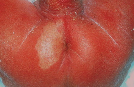
Perianal ulcerated hemangioma. The diagnosis is easier when observing the ischemic precursor

Downloads
DOI:
https://doi.org/10.26326/2281-9649.28.2.1849How to Cite
Abstract
Infantile hemangioma can ulcerate for various reasons, especially in the diaper region where there are particular conditions of humidity and temperature that macerate the skin and make it more susceptible to ulceration. The ulceration may be favored by ischemia which is currently considered an important pathogenetic factor in hemangioma. A finding supporting the role of ischemia in the pathogenesis of hemangioma is the initial clinical aspect of neonatal hemangioma precursors. These precursors may present as a pink spot or as telangiectases within an ischemic area or more rarely as an ischemic patch. These are different aspects of the same lesion that starts out as an ischemic patch, in which telangiectases are then formed; the latter then get confluent to give a flat pink spot; on the flat spot then proliferative papules are formed with the typical strawberry appearance (3, 4). We do not know if all the hemangiomas pass through these intermediate phases, but we have certainly documented in some cases these different passage phases. When we clinically observe these early stages of hemangioma, in most cases we experience poor growth, so many Authors talk about abortive hemangiomas or minimal growth hemangiomas.
In the literature there are some cases of neonatal perianal ulcers (1, 2, 5, 6) in which the histological appearance, the positivity of Glut1 (2) and sometimes the presence of peripheral telangiectasies lead to the diagnosis of hemangioma. In none of these cases is mentioned an earlier ischemic phase in the neonatal period as in our case. However, it is possible that this phase has gone unnoticed, unlike what we had the opportunity to document.
