Microvenular hemangioma.
Downloads
How to Cite
Bonifazi E., Lastilla G., Tataranni M., Maiorano E. 2018. Microvenular hemangioma. Eur. J. Pediat. Dermatol. 7 (1):33-8.
pp. 33-8
Abstract
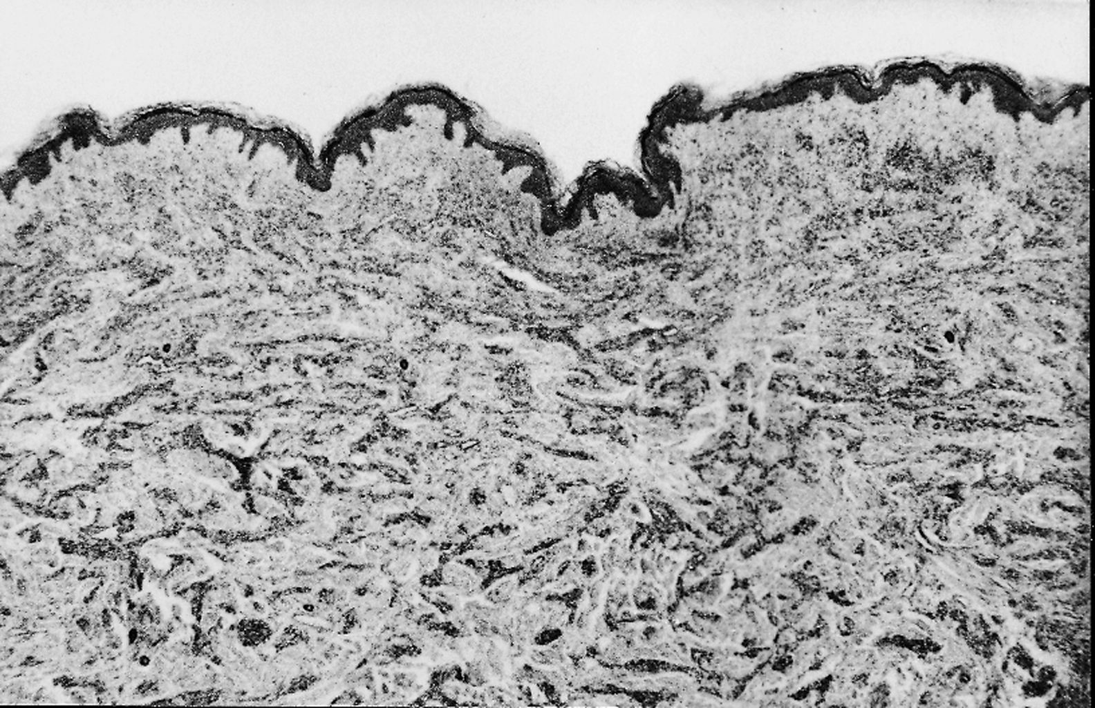
This study was aimed at evaluating the relationships between microvenular hemangioma and microcapillary hemangioma. A case of microvenular hemangioma in a 46-year-old woman is reported. The tumor mostly consisted of parallely oriented vessels composed of a three-layered wall with periluminal von Willebrand Factor-immunoreactive endothelial cells surrounded by muscle cells with smooth muscle actin immunoreactivity and by an outer layer of Collagen type IV-positive collagen. The histological and immunohistochemical findings of the present case confirm the venular nature of the majority of neoplastic vessels, supporting a reliable separation of this tumor from microcapillary hemangioma.
Keywords
Microvenular hemangioma, Microcapillary hemangioma
