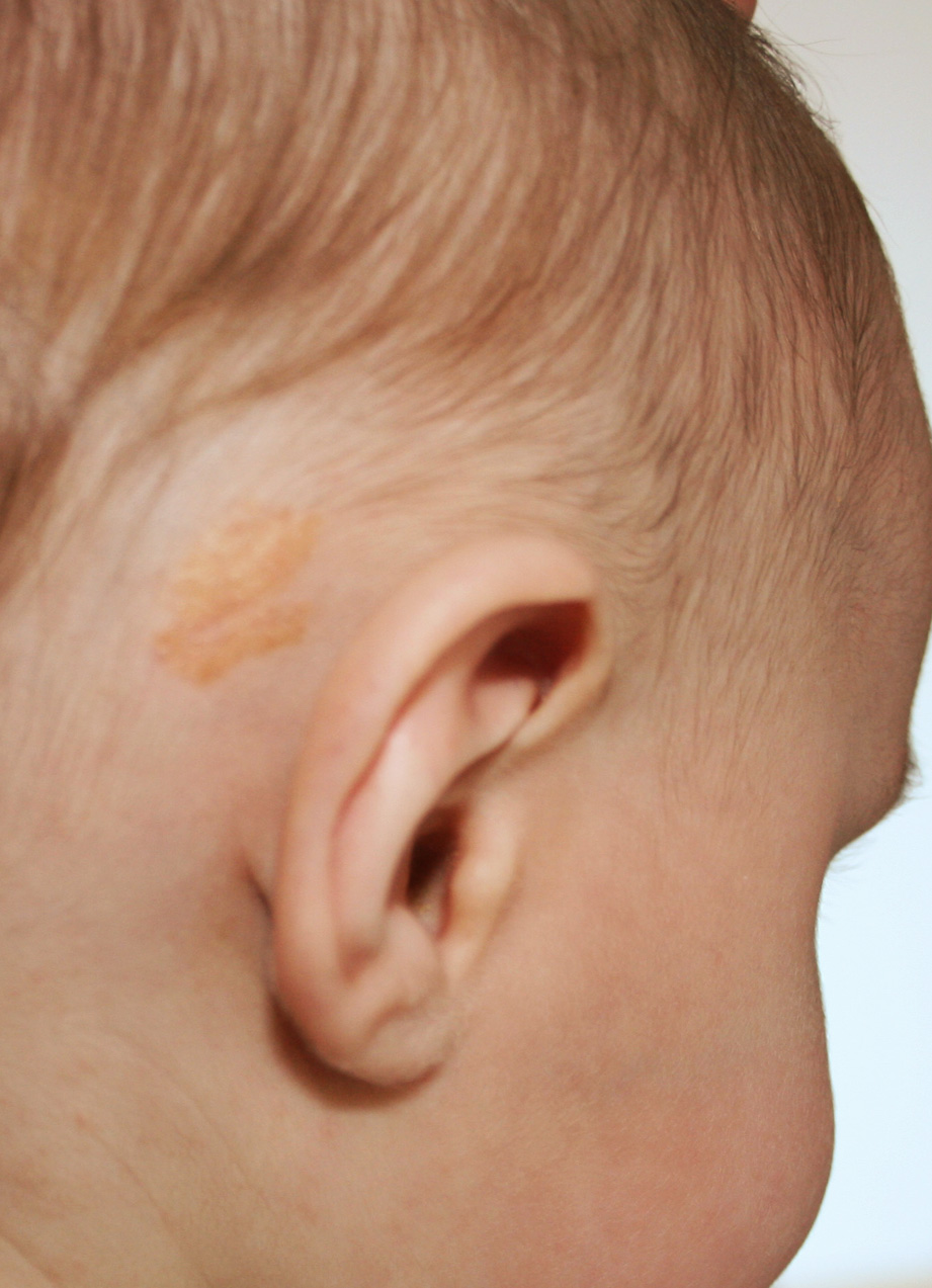
Chromatic variations in sebaceous nevus.

Downloads
DOI:
https://doi.org/10.26326/2281-9649.27.4.1502How to Cite
Abstract
The differential diagnosis of sebaceous nevus from aplasia cutis is sometimes difficult initially: the granular surface in SN and the absence of hair follicles in aplasia cutis are diriment. The incidence of neoplasms on SN was debated: a large report (2) in 596 cases of SN found basal cell carcinoma in 0.8% of cases and benign tumors in 13.6% on average in the 5th decade. More recently the dermoscopic findings of SN and their correlation with histological findings were reported (1). In the child prevail yellow lobules with a cobblestone appearance, histologically corresponding to hyperplastic sebaceous glands. In adults, a cerebriform appearance prevails due to the alternation of furrows and ridges corresponding to epidermal hyperplasia. In the actual case the darkening of SN may be related to thickened horny layer and the black dots surrounded by yellow halo to comedo-like openings surrounded by sebaceous glands.
