Longitudinal melanonychia in childhood.
Downloads
DOI:
https://doi.org/10.26326/2281-9649.27.4.1488How to Cite
Abstract
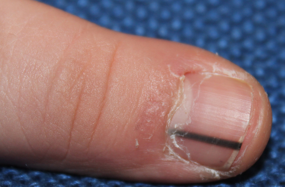
There are in the literature 11 cases of primary melanoma of the nail matrix presenting as longitudinal melanonychia (LM) in children. Asian or South American children with a dark phototype were mainly affected - 8 cases -, more rarely - 3 cases - Caucasian children with a fair-skinned phototype were involved and all of them were Italian. The fingers were involved in 8 cases, with prevalence of the 5th finger - 3/8 cases -, whereas the big toe was affected in 3 cases. In all cases the histological diagnosis was melanoma in situ; however, the differential diagnosis from benign melanocytic hyperplasia was almost always difficult. The clinical and dermoscopic signs allowing the differential diagnosis between malignant and benign LM in the adult are less valid in children. Moreover, also due to the anatomical and physiological peculiarities of the melanocytes of the nail matrix, there are no reliable histological criteria for this differential diagnosis. This is why longitudinal melanonychia in childhood should be very carefully monitored clinically, performing a biopsy of the nail matrix in case of rapid and persistent evolution of the lesion.
