The evolution of histopathological changes in dermatitis artefacta.
Downloads
DOI:
https://doi.org/10.26326/2281-9649.27.4.1485How to Cite
Abstract
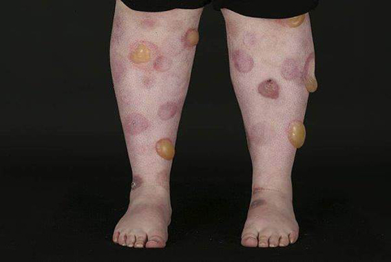
We present a case of a 17 year old female with a decade long history of a blistering skin disorder. She had been extensively investigated and was empirically being managed for a possible diagnosis of bullous lupus erythematosus. On presentation she had a widespread striking bullous eruption with relative sparing of her back. She was on multiple immunosuppressive medications and opiates for pain relief. Previous histopathology specimens were re-examined. One specimen showed a subepidermal blister mainly with fibrin and a neutrophilic inflammatory cell infiltrate. Two specimens showed cell poor subepidermal blisters. The differential diagnosis included bullous lupus erythematosus, dermatitis herpetiformis, linear IgA disease, erythema multiforme and toxic epidermal necrolysis, amongst others. However, given the negative immunofluorescence studies and the clinical history, these differential diagnoses seemed unlikely. It was felt that the appearances in all the biopsies most likely represented the same process in different stages of evolution. Overall these findings were suggestive of a blister induced by a physical agent, possibly a suction blister. A diagnosis of dermatitis artefacta was made and all immunosuppressive medications were discontinued. Our case highlights the importance of careful clinicopathological correlation in order to avoid errors in diagnosis and unnecessary treatments. It also demonstrates the potential histopathological heterogeneity that can be observed when multiple biopsies are taken at different times in the course of the presentation and that this is likely to represent the same process at different stages of evolution.
