Perilesional dermatitis after pyoderma
Downloads
DOI:
https://doi.org/10.26326/2281-9649.27.3.1471How to Cite
Abstract
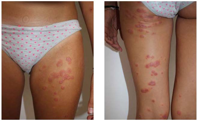
In 2008, we described two children who after a banal bullous pyoderma showed around the hypomelanic outcomes of the disease erythematous-infiltrative lesions in annular plaques that regressed in a dozen days and did not recur. The morphology of the lesions recalled erythema multiforme and for this reason we talked about perilesional dermatitis reminiscent of erythema multiforme (2). However, the asymmetric distribution of the polymorphous lesions occurring at the periphery of pyoderma lesion outcomes is not at all related with erythema multiforme, recalling, however, for localization and time of onset of allergic contact dermatitis, particularly experimental sensitization, mosquito hypersensitivity and allergic reactions to tattoos. In experimental contact sensitization when a concentrated solution of an allergen is applied on the skin after a period of 7 to 10 days, which is necessary because the immune system produces a lymphocyte clone capable of recognizing and reacting with the allergen, there is a flare up, i.e. the re inflammation of the skin site on which the allergen has been applied. The flare up is attributed to the encounter of circulating lymphocytes that are sensitized to the allergen with residues of the same allergen remaining in the site of the first application. From the moment of flare up, much diluted solutions of the allergen applied at any point on the
skin surface cause an allergic contact dermatitis (4). Something like that occurs in hypersensitivity to mosquito that is due to associated immediate and delayed allergic reactions (3). Often, people with mosquito hypersensitivity report that after a single mosquito puncture all the sites of the previous bites are re-activated. Moreover, we also document pediatric mosquito hypersensitivity cases that start in winter (1) in the absence of mosquitoes. We attributed these cases to a recent onset of mosquito hypersensitivity in those children: when this event occurs in winter hypersensitivity reactions appear out of season due to the persistence of mosquito allergens in the skin of previous summer bites; the latter had not been clinically noted because at that time there was not yet hypersensitivity to mosquitoes in those children. We hypothesize that the same pathogenetic mechanism works in case of skin infections, for example due to Staphylococcus aureus, whose allergenic ability is well known, especially in the atopic subject: during the infection the subject could become sensitized to bacterial allergens and in the remission phase the circulating sensitized lymphocyte clone, coming in contact with allergenic residua present at the infection site may result in the infiltrative erythematous lesions we have observed. The clinical lesions are not weals, leading to the exclusion of an IgE-mediated reaction; they are not even eczematous as in allergic contact dermatitis. However, they are reminiscent of infiltrative erythematous reactions in subjects with delayed allergic reactions to red tattoo (5).
