Molluscum contagiosum with unexpected outcome.
Downloads
DOI:
https://doi.org/10.26326/2281-9649.26.4.1293How to Cite
Simeone D., Bonifazi E. 2016. Molluscum contagiosum with unexpected outcome. Eur. J. Pediat. Dermatol. 26 (4): 248. 10.26326/2281-9649.26.4.1293.
pp. 248
Abstract
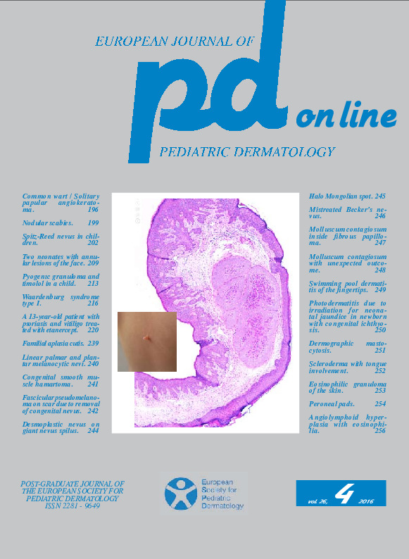
The epidermal proliferation induced by molluscum contagiosum virus gives rise to a pear-shaped epidermis lobule (Fig. 2) with the base going down in the dermis; a pear-shaped lobule corresponds to an element of molluscum contagiosum.There may be more lobules separated and isolated from each other or they can be close to each other, separated by thinned dermal papillae reduced to thin fibrous septa, giving rise to plaques composed of many agminate lobules (2, 3).
In the giant molluscum contagiosum (1) many lobules or molluscum contagiosum elements can be close to each other to form a 2 cm in size nodule (Fig. 3).
We hypothesize that the mammilated mass of our case was due to an endophytic development of several close to each other but non confluent elements; the pressure exerted on the side walls of the mass would have resulted in the release of the same.
Keywords
molluscum contagiosum
