Hypopigmentation due to clobetasol.
Downloads
How to Cite
Alaibach M., Bonifazi E. 2014. Hypopigmentation due to clobetasol. Eur. J. Pediat. Dermatol. 24 (2): 125.
pp. 125
Abstract
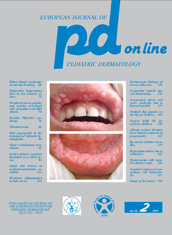
Case 1. An 18-year-old boy after a diagnosis of cutaneous plaque scleroderma had been treated for 5 years with systemic and topical corticosteroids (clobetasol propionate) and methotrexate in a Rheumatology Department. His history began when aged 13 with a hypertrichotic and hypermelanic patch on the right shoulder. At that time, a skin biopsy was compatible with scleroderma or lichen sclerosus. 5 years later a dermatologist advanced the suspicion of a nevus lesion. The dermatological examination (Fig. 1) showed a normal skin texture with patches of hypertrichosis and hyperpigmentation surrounded by hypopigmented areas. The history and clinical features led to diagnose Becker's nevus with hypopigmentation due to clobetasol. Case 2. A 4-year-old little girl with segmental vitiligo of the back was recommended to apply topical clobetasol only on the lateral patch of vitiligo. At control the patient showed hypopigmentation (arrows) surrounding the treated vitiligo patch (Fig. 2), leading to diagnose hypopigmentation due to clobetasol.Keywords
Hypopigmentation, Clobetasol
